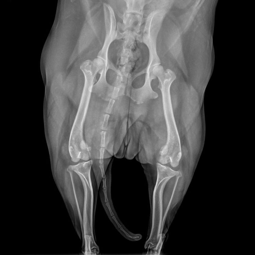
Medial Patellar Luxation (MPL) Repair surgery corrects instability of the kneecap, which can move out of its normal position. The surgeon deepens the groove in the femur and realigns the attachment point of the patellar ligament.
This procedure is commonly recommended for dogs experiencing limping, intermittent skipping, or discomfort due to kneecap instability.
We focus on restoring proper alignment and function of the joint to improve comfort, mobility, and long-term limb health.
Surgeons use a 1-to-4 scale to determine the severity and necessity of surgery:
Grading Scale
Grade
1
The kneecap can be manually pushed out but pops back in immediately. These pets rarely show symptoms and often don’t need surgery.
Grade
2
The kneecap slips out spontaneously during activity, causing a “skipping” gait. The pet may kick the leg out to “snap” it back in. Surgery is often recommended to prevent long-term wear.
Grade
3
The kneecap is out of the groove most of the time but can be manually pushed back in. These pets usually have a consistent limp.
Grade
4
The kneecap is permanently dislocated and cannot be manually replaced. This often involves significant bony deformities and requires complex reconstructive surgery.
While it is most famous in toy breeds like Chihuahuas and Yorkies, it also affects larger dogs and is often a result of congenital skeletal misalignments.
Surgical Techniques
Because MPL is usually caused by multiple alignment issues, surgeons often combine several procedures to fix it:
Trochleoplasty: Deepening the shallow groove (trochlea) in the femur so the kneecap has a “taller” wall to sit behind.
Tibial Tuberosity Transposition (TTT): The bony point where the patellar tendon attaches to the tibia is cut and moved to align it directly under the groove. It is then secured with orthopedic pins.
Soft Tissue Imbrication: Tightening the loose tissues on the outside of the knee and releasing the tight ones on the inside to help “center” the kneecap.
Expected Outcomes & Success Rates
Success Rate: Approximately 90% of pets see a dramatic improvement in mobility and comfort after surgery.
Healing Timeline: Strict confinement (crate or small room) is required for 6 to 8 weeks to allow the bone to fuse back together where it was moved.
Rehab: Professional physical therapy, including underwater treadmills and range-of-motion exercises, is highly recommended to prevent joint stiffness and rebuild muscle.